Abstract
Alopecia areata (AA) is a common autoimmune disease causing non-scarring hair loss. While typically presenting with round or oval patches, this report describes two unique cases of AA exhibiting an annular pattern, a previously unreported presentation in English literature.
Introduction
AA affects 1.7% of the population at some point in their lives and is characterized by T-cell mediated hair follicle destruction. Clinical presentations vary, with the most common being round or oval patches of hair loss. Here, we report two unusual cases of AA presenting with an annular pattern.
Case Presentation
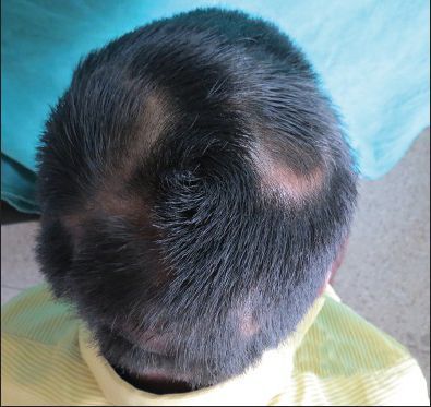
A 25-year-old male presented with a two-month history of asymptomatic, annular hair loss on the scalp. There was no family history of similar conditions, no history of medication use, and no scalp trauma. Examination revealed multiple, well-defined, annular patches of non-scarring alopecia ranging from 2x3 cm to 4x4 cm, primarily located on the parietal and occipital scalp (Figure 1a). Scalp skin was smooth with no associated abnormalities. Systemic examination was normal.
Investigations
Potassium hydroxide (KOH) examination and fungal culture ruled out fungal infection. Histopathological examination of a hairless region showed sparse perifollicular lymphocytic infiltrate with vellus hair follicles in the mid-dermis, consistent with AA (Figure 1b).
Diagnosis
Based on clinical presentation and histopathology, the diagnosis of annular AA was established.
Treatment and Follow-Up
The patient received intralesional triamcinolone acetonide injections (5 mg/ml) every four weeks. After three months of follow-up (3 injections), he showed significant improvement in hair regrowth (Figure 1c).
Discussion
AA typically presents with round or oval patches of hair loss. This report highlights two unique cases with an annular pattern, previously unreported in English literature. While the course of treatment and response remained consistent with standard AA management, this case demonstrates the diverse morphological presentations AA can exhibit.
Conclusion
This report broadens our understanding of AA's clinical presentation. While the most common patterns are round or oval, AA can manifest in various forms, including the previously unreported annular pattern. Recognizing these variations is crucial for accurate diagnosis and appropriate management.
Figures
Figure 1a: Annular patches of hair loss on the scalp.
Figure 1b: Histopathological examination showing sparse lymphocytic infiltrate and vellus hair follicles.
Figure 1c: Improvement in hair regrowth after treatment.
Diagnostics
| Date | Type | Value | Unit |
|---|


.png)


2 Comments